

When is testing SERUM for Histoplasma antigen indicated?
Case Example
Lacey, a 3-year-old, spayed female Chihuahua was presented for chronic large-bowel diarrhea and intermittent anorexia of 2 years duration. Clinical signs had not improved with tylosin and hypoallergenic diet. Routine lab work showed mild hypoalbuminemia (2.4 g/dL; RI 2.7-4.4), mature neutrophilia (23,306 / mcL; RI 2060-10600), monocytosis (3252 / mcL; RI 0-840), and lymphopenia (542 / mcL; RI 690-4500). Serum cobalamin, folate, cPLI, and TLI concentrations were normal. Fecal flotation for intestinal parasites and fecal PCR for Herterobilharzia were negative. Serum IBD panel showed elevated anti-porin IgA, anti-calprotectin IgA, and anti-giladin IgA, suggestive of inflammatory bowel disease. Abdominal ultrasound showed a thickened colonic wall with segmental loss of layering and mesenteric lymphadenopathy (Figure 1). Urine was negative for antigen with the MVista® Histoplasma Antigen Quantitative EIA.
![]()
Figure 1: Abdominal ultrasound showing thickened colonic wall with loss of normal layering (arrow).
Based on the presumptive diagnosis of inflammatory bowel disease, prednisone was prescribed. Short term clinical improvement was minimal. Due to the continued suspicion for GI histoplasmosis, serum was tested for antigen by the MVista® Histoplasma Antigen Quantitative EIA (test code 310) and for antibodies by the MVista® Canine Histoplasma IgG Antibody EIA (test code 327). Histoplasma antigen was detected in serum (1.2 ng/ml; RI=none) as were anti-Histoplasma IgG antibodies (>80 EU; RI <8). Considering Lacey’s clinical findings, either test result is sufficient to make the diagnosis of GI histoplasmosis.
Discussion
Lacey’s case is a good example of when to test serum for Histoplasma antigen. When urine is tested for antigen with the MVista® Histo Antigen EIA it is highly sensitive (89% dog and 94% cat) for histoplasmosis [1
In cats, localized histoplasmosis most often affects the eyes and bone/joints and less commonly the skin and GI tract. In a review of 183 samples submitted to MiraVista Diagnostics, approximately 3% of urine samples were negative when tested or Histoplasma antigen while serum, submitted at the same time, was positive. This data suggests that if only urine is tested, the diagnosis will be missed in at least 3% of samples. This number does not include cases where both urine and serum antigen tests are negative, which is estimated to occur in about 5% of cases. In that case, testing for anti-Histoplasma antibodies with the MVista® Histoplasma IgG Antibody EIA is indicated. In Lacey’s case, the Histoplasma IgG antibody concentration was very high (>80 EU).
Lacey is a classic example of GI histoplasmosis. Large bowel involvement is almost always present and often diarrhea contains frank blood or mucous or both. Straining and urgency might also be present. Affecting the most distal GI tract, proctitis is also relatively common. Routine lab work can be normal or have changes consistent with chronic inflammation. Abdominal ultrasound generally reveals a thickened colonic wall with a loss of normal layering. Local mesenteric lymph nodes can be enlarged. Cytology of FNA of mesenteric lymph nodes will often show evidence of antigenic stimulation, but not Histoplasma yeasts. A rectal scrape, where rectal mucosal cells are digitally scraped off and rolled onto a slide, might reveal Histoplasma yeast (Figure 2). As with Lacey’s case, many dogs have clinical signs for months or even years before a definitive diagnosis is made. During that time, there is usually short term and incomplete response to multiple empiric treatments.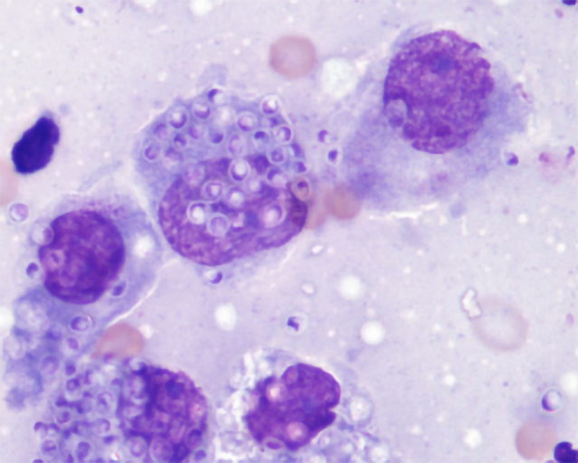
Figure 2: Histoplasma yeasts seen mostly within macrophages. Photo courtesy of Dr. Jim Meinkoth.
Other common diagnostic rule-outs for a case similar to Lacey’s include intestinal neoplasia, such as lymphoma, and inflammatory bowel disease. Other endemic mycoses such as blastomycosis, coccidioidomycosis and cryptococcosis much less commonly affect the GI tract. Pythiosis more commonly affects the stomach or small bowel. One additional important aspect of Lacey’s case includes the positive results on the serum IBD panel. In enzootic areas, GI histoplasmosis must be ruled-out before an IBD panel can be used to make a presumptive diagnosis of inflammatory bowel disease.
REFERENCES:
- Rothenburg L, Hanzlicek AS, Payton ME. A monoclonal antibody-based urine Histoplasma antigen enzyme immunoassay (IMMY(R)) for the diagnosis of histoplasmosis in cats. J Vet Intern Med 2019;33:603-610.
- Cunningham L, Cook A, Hanzlicek A, et al. Sensitivity and Specificity of Histoplasma Antigen Detection by Enzyme Immunoassay. J Am Anim Hosp Assoc 2015;51:306-310.
- Cook AK, Cunningham LY, Cowell AK, et al. Clinical evaluation of urine Histoplasma capsulatum antigen measurement in cats with suspected disseminated histoplasmosis. J Feline Med Surg 2012;14:512-515.
- Hanzlicek AS, Meinkoth JH, Renschler JS, et al. Antigen Concentrations as an Indicator of Clinical Remission and Disease Relapse in Cats with Histoplasmosis. J Vet Intern Med 2016;30:1065-1073.
- Smith KM, Strom AR, Gilmour MA, et al. Utility of antigen testing for the diagnosis of ocular histoplasmosis in four cats: a case series and literature review. J Feline Med Surg 2017;19:1110-1118.
- Fielder SE, Meinkoth JH, Rizzi TE, et al. Feline histoplasmosis presenting with bone and joint involvement: clinical and diagnostic findings in 25 cats. J Feline Med Surg 2019;21:887-892.